Biological pregnancy test
Detecting a hormone through its effect on an animal
Contents
- 1Urine sample posted in its box
- 2Tank with a female clawed frog
- 3Microscope
- 4Rack of test tubes
- 5Register of results
A biological pregnancy test detected pregnancy from the effect a woman's urine had on a laboratory animal. The urine of a pregnant woman carries a hormone made by the placenta, and that hormone acts on the ovaries or testes of a mouse, a rabbit, a frog or a toad. Selmar Aschheim and Bernhard Zondek described the reaction in Berlin in 1927. For several decades specialist laboratories ran these tests at a doctor's request, until tests based on antibodies made the animals unnecessary.
What it is
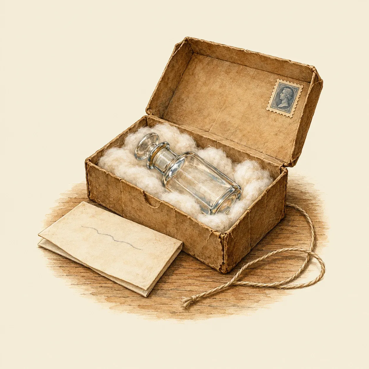 Doctors sent the sample by post and received the answer by post.
A biological pregnancy test answered one question: does this urine contain the hormone of pregnancy? At the time no chemical method could find the hormone in a sample. A laboratory gave the urine to an animal and watched for a change that this hormone causes. From the late 1920s to the 1960s pregnancies were confirmed in this way by specialist laboratories, in hospitals and in a few university departments. In Britain the best-known service was run at the University of Edinburgh by the geneticist Francis Crew, in his department of animal breeding research. Doctors posted a small bottle of morning urine with a few notes on the patient, and the answer came back by post. Crew turned down requests sent directly by women: the test went through a doctor, who received the result and passed it on.
How it works
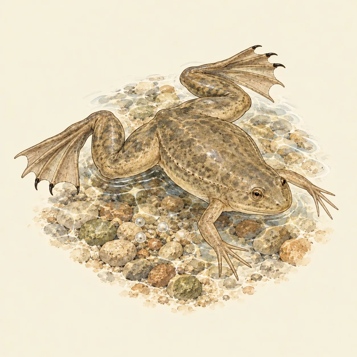 The clawed frog lays eggs within hours when the hormone is present, and can be used again.
From the first weeks of pregnancy the placenta produces a hormone, human chorionic gonadotropin or hCG, and the kidneys pass part of it into the urine. This hormone acts on the ovaries in the same way as a hormone of the pituitary gland, the small gland under the brain that makes ovaries mature. An immature female mouse has inactive ovaries. When it received urine containing hCG, its ovaries matured within a few days, and the laboratory recognised the change under the microscope. Urine without the hormone left the ovaries unchanged. Rabbits react the same way and faster.
Amphibians made the reading easier. A female African clawed frog, Xenopus laevis, lays eggs within hours when she receives the hormone, so the result can be seen in the tank, and the same frog can be used for later tests. A male toad responds by releasing sperm cells, which the laboratory found in a drop of its urine under the microscope. Since the hormone comes from the placenta, the test detects living placental tissue and not the embryo itself.
History and evolution
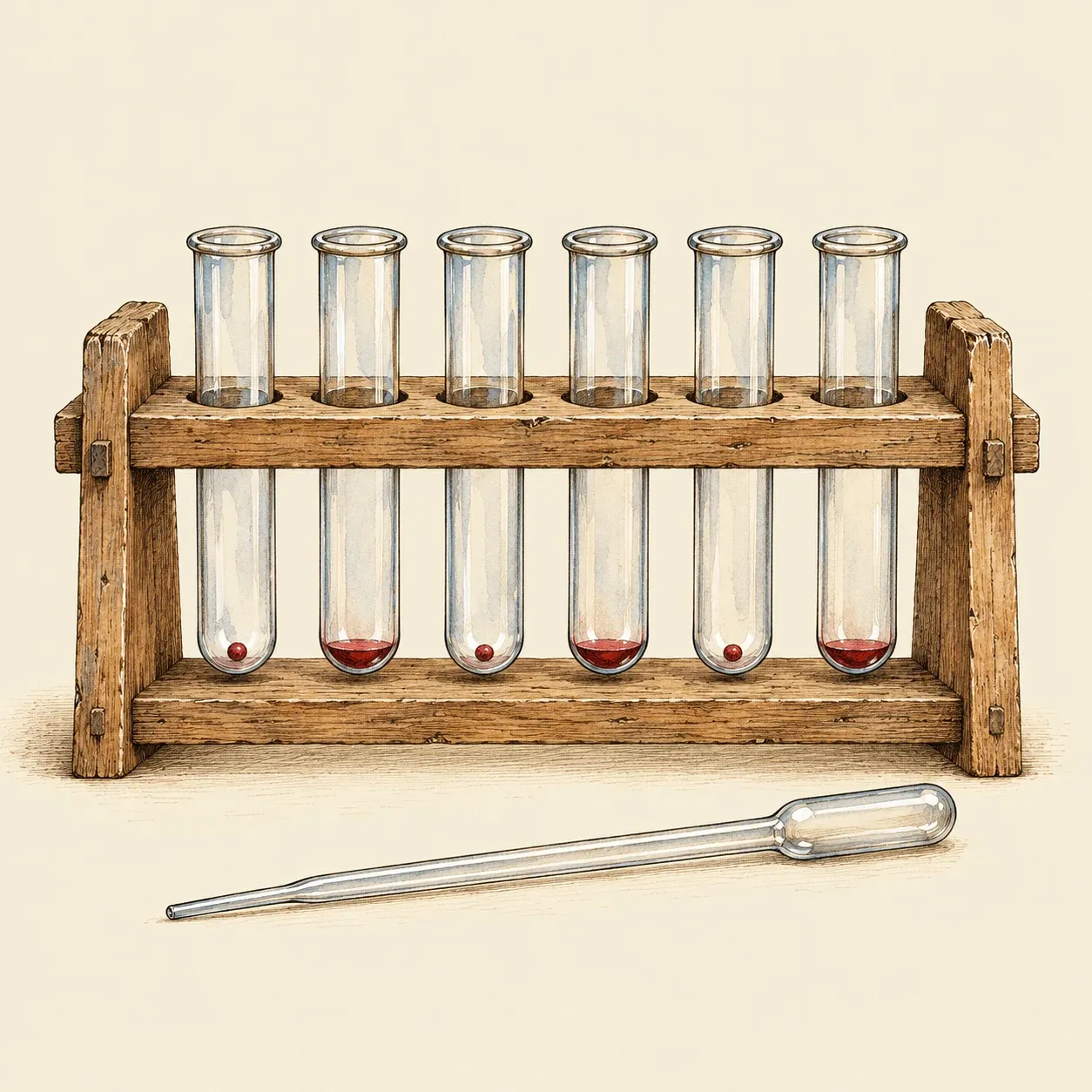 In the antibody test, red cells that settle as a ring or a dot give the answer without any animal.
In 1927 Selmar Aschheim and Bernhard Zondek, gynaecologists in Berlin, reported that the urine of pregnant women acted on immature mice like the pituitary gland itself. They turned the finding into a diagnostic method published in 1928, and an editorial in the Lancet soon presented it to British doctors. Crew’s Edinburgh station then began testing samples posted by doctors. In 1931 Maurice Friedman and Maxwell Lapham in Philadelphia used adult female rabbits and brought the wait down from about five days to one or two. In Cape Town, Lancelot Hogben had shown that the clawed frog lays eggs in response to the pituitary hormone. His former students and other researchers proposed the frog as a pregnancy test, and Hogben pointed out to Crew an animal that ovulates visibly and can be used several times. Frogs later largely replaced mice and rabbits. In Buenos Aires Carlos Galli Mainini published a test on male toads.
Animals left the laboratory with antibodies, the proteins of the immune system that recognise one precise substance, the same defences that vaccination trains. Leif Wide and Carl Gemzell in Sweden published a test in 1960 that mixed the urine with antibodies against hCG and with red blood cells coated with the hormone. If the urine contained hCG, it occupied the antibodies, and the red cells settled in a neat ring instead of clumping. In 1972 Judith Vaitukaitis and her colleagues in the United States described an assay that told hCG apart from the similar pituitary hormone LH. Kits for use at home went on sale in the 1970s. Monoclonal antibodies made the tests more sensitive in the 1980s, and in 1988 a one-step stick gave its reading in three minutes. The pregnancy tests used today are validated medical devices that need no live animal. The microscope that once read the mouse and toad reactions is still used in many other laboratory tests.